Skip to content
PVG INTERNATIONALPVG INTERNATIONAL
    • Mobile: +91-9466446612 +91-9728497962
    • E-Mail: info@pvginternationals.com info@narayanienterprises.com
  • Cart

    No products in the cart.

  • Home
  • About Us
  • Certifications
  • Products
  • Contact
  • Blog
Add to wishlist
Awaiting product image
Home / Anatomical Models

YM-195 MODEL OF SEED GERMINATION

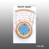
  • Model of dicot root
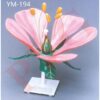
  • DICOT FLOWER MODEL Flowers represent an important adaptation for plants, as the different shapes and colors are used to attract different pollinating species of insects and other animals. The colorful flowers characteristic of dicots consist of a specific number of sepals, or petals. In dicots, the number of petals occurs in groups of four or five . PVG’s Typical Dicot Flower Model is enlarged, Educational, 3 D plastic, unbreakable model, this model is for  students to easily study the floral components and the reproductive functions in flowers. Model can be disassembled to reveal the pollen grains inside, showing the process of angiosperm pollination and fertilization . Important structures are numbered on the flower model and corresponds to an identification key. THIS Model includes structures of Flower like receptacle, petal, sepal, gynoecium (stigma and style), male stamen (anther and filament), pollen, pollen tube etc. Petals, sepals and  stamens are detachable. Ovary is dissected.  Hand painted each part in different color, to deepen the understanding of the structure of the dicotyledonous flower. Sepals are colored green, petals, pink, ovary green, and anthers with pollen tubes in yellow color. This Model is Botanical teaching aid for Dicot plants. Saves learning time and improve learning efficiency This is a perfect model for teachers to make students to understand the Anatomy of Flower in Botanical classes.    

Comparing germination in corn and bean. On base size 16×12”

SKU: YM-195 SEED GERMINATION Category: Anatomical Models
  • Model of dicot root
  • DICOT FLOWER MODEL Flowers represent an important adaptation for plants, as the different shapes and colors are used to attract different pollinating species of insects and other animals. The colorful flowers characteristic of dicots consist of a specific number of sepals, or petals. In dicots, the number of petals occurs in groups of four or five . PVG’s Typical Dicot Flower Model is enlarged, Educational, 3 D plastic, unbreakable model, this model is for  students to easily study the floral components and the reproductive functions in flowers. Model can be disassembled to reveal the pollen grains inside, showing the process of angiosperm pollination and fertilization . Important structures are numbered on the flower model and corresponds to an identification key. THIS Model includes structures of Flower like receptacle, petal, sepal, gynoecium (stigma and style), male stamen (anther and filament), pollen, pollen tube etc. Petals, sepals and  stamens are detachable. Ovary is dissected.  Hand painted each part in different color, to deepen the understanding of the structure of the dicotyledonous flower. Sepals are colored green, petals, pink, ovary green, and anthers with pollen tubes in yellow color. This Model is Botanical teaching aid for Dicot plants. Saves learning time and improve learning efficiency This is a perfect model for teachers to make students to understand the Anatomy of Flower in Botanical classes.    
Categories
  • Chemistry - General & Chemical Laboratory Apparatus
  • Laboratory Glassware
    • Distillation Assembly & Rotary Evaporators
    • Laboratory Glassware section -1
    • Interchangeable Laboratory Glassware
    • Organic Chemistry Set
    • Clevenger, Dean & Stark
    • Chromatography Columns & Gas Analysis
    • Clinical Laboratoy Glassware
    • Dairy & Milk Testing
    • Hydrometers
    • Thermometers
    • Quartz Glassware
    • Sintered Glasswae
    • Reaction Vesssel
    • Volumetric Glasswares
  • Anatomical Models
  • Biological Apparatus & Material
  • Microscopes & Accessories
  • Physics & Electronic Laboratory Apparatus
  • Pharmacy Apparatus
  • Earth Sciences
  • Plasticware
  • Uncategories
  • Agriculture Equipment
  • Surgical Instruments & Hospital Equipment
  • Chemicals
  • Material Testing
Recently Viewed
  • Economical model, showing complete circulatory system i.e. heart, arteries, veins etc. Size 60 x 25 x 35 mm. YM-130 MODEL OF HUMAN CIRCULATORY SYSTEM (ECONOMICAL)
  • For artificial and natural gas. Adjustable orifice with regulated gas supply. Total Height 240 mm., diameter of grid top 38 mm. Also available with height 175 mm. and grid top 25 mm PM-202 BURNER HIGH TEMPERATURE
  • Comprises a borosilicate glass U tube with 225 mm. height, with each limb graduated 0-120 x 1 mm. Bend fitted with a filling tube and stopcock. Two platinum electrodes 14 mm. diameter can be adjusted at any position by locking PM-199 BURNER TECLU
  • Reviews (0)

Reviews

There are no reviews yet.

Be the first to review “YM-195 MODEL OF SEED GERMINATION” Cancel reply

Related products

Excellent Educational model to teach Medical students the various parts of skull, its anatomy.Very helpful for Biology students to Understand the Anatomy of Skull. Superior model,three part, fibre glass, with full dentition and all other details. The calvaria is uncut and the lower jaw is spring held.
Add to wishlist
Quick View

Anatomical Models

YM-09 HUMAN SKULL

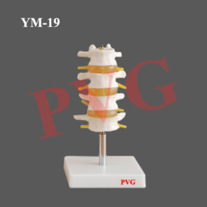
YM-12 VERTEBRAL COLOUMN WITH PELVIS THIS MODEL SHOWS ALL SIGNIFICANT FEATURES OF EACH VERTEBRA, , A  HERNIATED DISC AND VERTEBRAL NOTCH ETC. SPECIAL FEATURES INCLUDE: FLEXIBLE 29" TALL VERTEBRAL COLUMN COMPLETE WITH PELVIS, SACRUM, OCCIPITAL BONE, HERNIATED LUMBAR  DISC. DELUXE CHROME STANDS 34" HIGH   YM-12 A VERTEBRAL COLOUMN WITH PELVIS THIS MODEL SHOWS ALL SIGNIFICANT FEATURES OF EACH VERTEBRA, INCLUDING SPINAL CORD, NERVE ROOTS, THE VERTEBRAL ARTERY, A  HERNIATED DISC AND VERTEBRAL NOTCH ETC. SPECIAL FEATURES INCLUDE: FLEXIBLE 29" TALL VERTEBRAL COLUMN COMPLETE WITH PELVIS, SACRUM, OCCIPITAL BONE, VERTEBRAL ARTERY, ALL NERVE BRANCHES AND HERNIATED LUMBAR DISC. DELUXE CHROME STANDS 34" HIGH
Add to wishlist
Quick View

Anatomical Models

YM-12 HUMAN VERTEBRAL COLUMN

This model shown the osteoporosis and blood vessel.This model shown the osteoporosis and blood vessel.
Add to wishlist
Quick View

Anatomical Models

YM-20 FEMUR BONE

YM- 41 FISH MODEL
Add to wishlist
Quick View

Anatomical Models

YM- 41 FISH MODEL

HAND JOINT WITH LIGAMENTS LIFE SIZE
Add to wishlist
Quick View

Anatomical Models

YM-16 HAND JOINT WITH LIGAMENTS LIFE SIZE

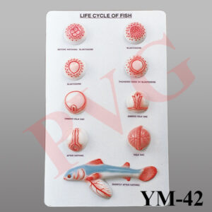
Set of nine stages from blastoderm to yolk sac and stage shortly after hatching. On base size 12 x 20”.
Add to wishlist
Quick View

Anatomical Models

YM-42 LIFE CYCLE OF FISH

KNEE FUNCTIONAL JOINT LIFE SIZE WITH MUSCLES
Add to wishlist
Quick View

Anatomical Models

YM-24 KNEE ANATOMY LIFE SIZE MODEL

Placeholder
Add to wishlist
Quick View

Anatomical Models

YM- 05 MODEL OF DISARTICULATED HUMAN SKELETON (200 Bones)

  • Home
  • About Us
  • Certifications
  • Products
  • Contact
  • Blog

Address Details

PVG International
# 6 - C, New Ram Nagar, Ambala Cantt - 133 001, Haryana, India.
# P.O. Box-113

Our Visitor

081869

Contact Details

  • info@pvginternationals.com
  • info@narayanienterprises.com
  • +91-9466446612
  • +91-9728497962
  • Phone: +91-171-2651006, 2653272
  • Fax: +91-171-2653259
Copyright 2026 © PVG INTERNATIONAL
  • Home
  • About Us
  • Certifications
  • Products
  • Contact
  • Blog
  • Login
    • Mobile: +91-9466446612 +91-9728497962
    • E-Mail: info@pvginternationals.com info@narayanienterprises.com

Login

captcha

Lost your password?

Register

Your personal data will be used to support your experience throughout this website, to manage access to your account, and for other purposes described in our privacy policy.

captcha