


MODEL OF ANIMAL MITOSIS
This model shows different phases in Cell Division.
This innovative model set is used to teach the Medical students mitotic division.
Nine phases of animal cell mitosis are depicted on individual removable models that are very much enlarged.
In each phase nucleus, centrioles, centrosome, chromatin, Chromosomes, spindle, aster and other main components are depicted properly to understand Through this model in The process of cell divison,
Each phase is painted properly using the vibrant colors allowing students to easily understand the different phases
Individual models are magnetic and can be arranged on whiteboards in your
The following phases of mitosis are depicted: interphase, prophase, early prometaphase, late prometaphase, metaphase, early anaphase, late anaphase, telophase, and cytokinesis.
Be the first to review “YM-30 ANIMAL MITOSIS SET”
Related products

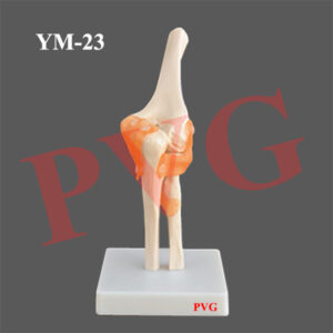
Anatomical Models

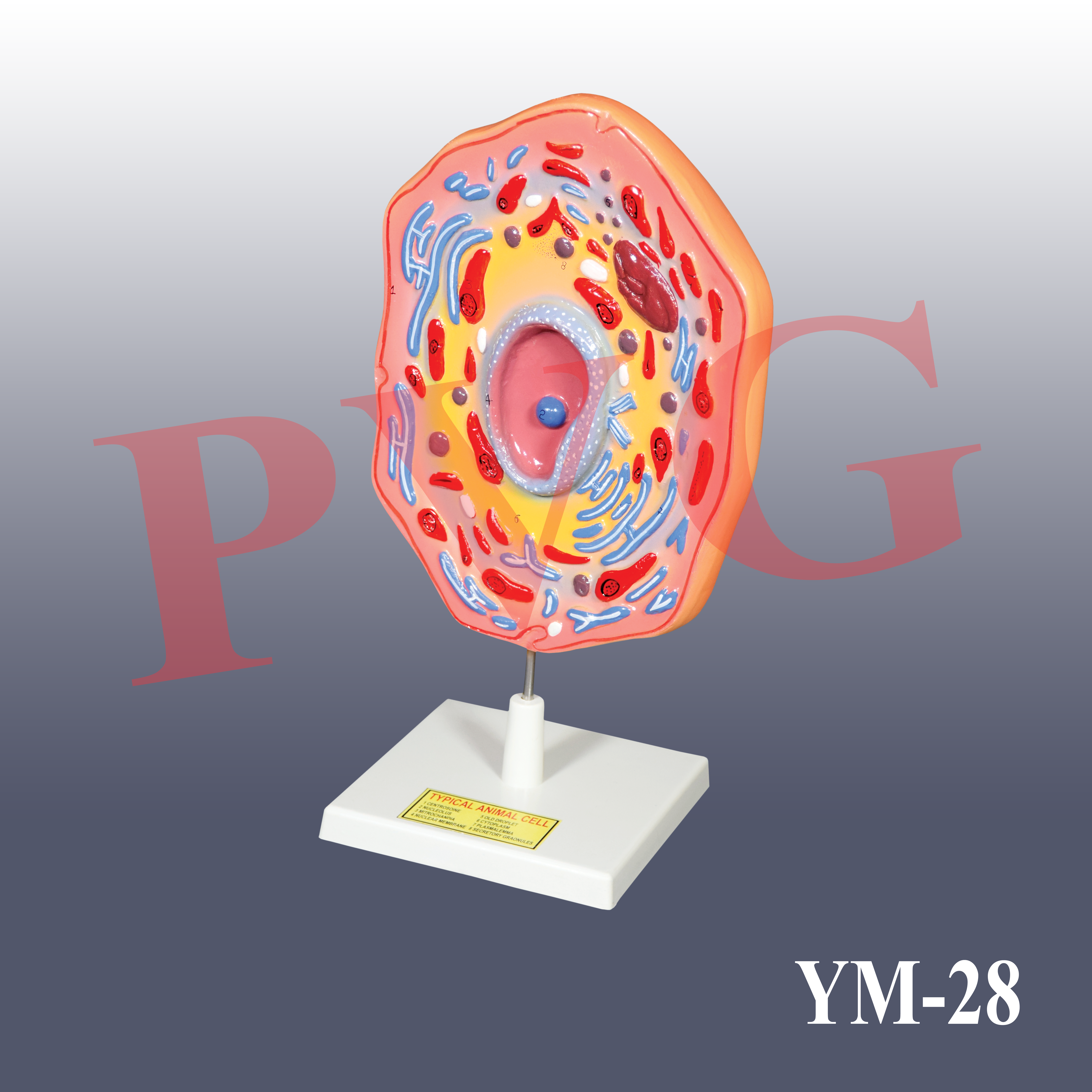
Anatomical Models

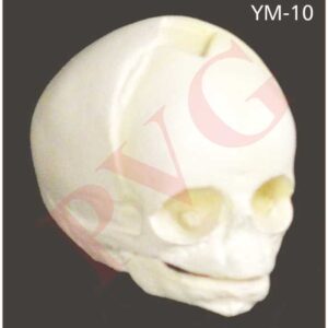
Anatomical Models
Anatomical Models
Anatomical Models

Anatomical Models

Anatomical Models
Anatomical Models
Reviews
There are no reviews yet.