
YM-191 LEAF TRANSVERSE SECTION DICOT (ECONOMICAL)
Economical model for students depicting the Dicot leaf structure
mounted on board with key card
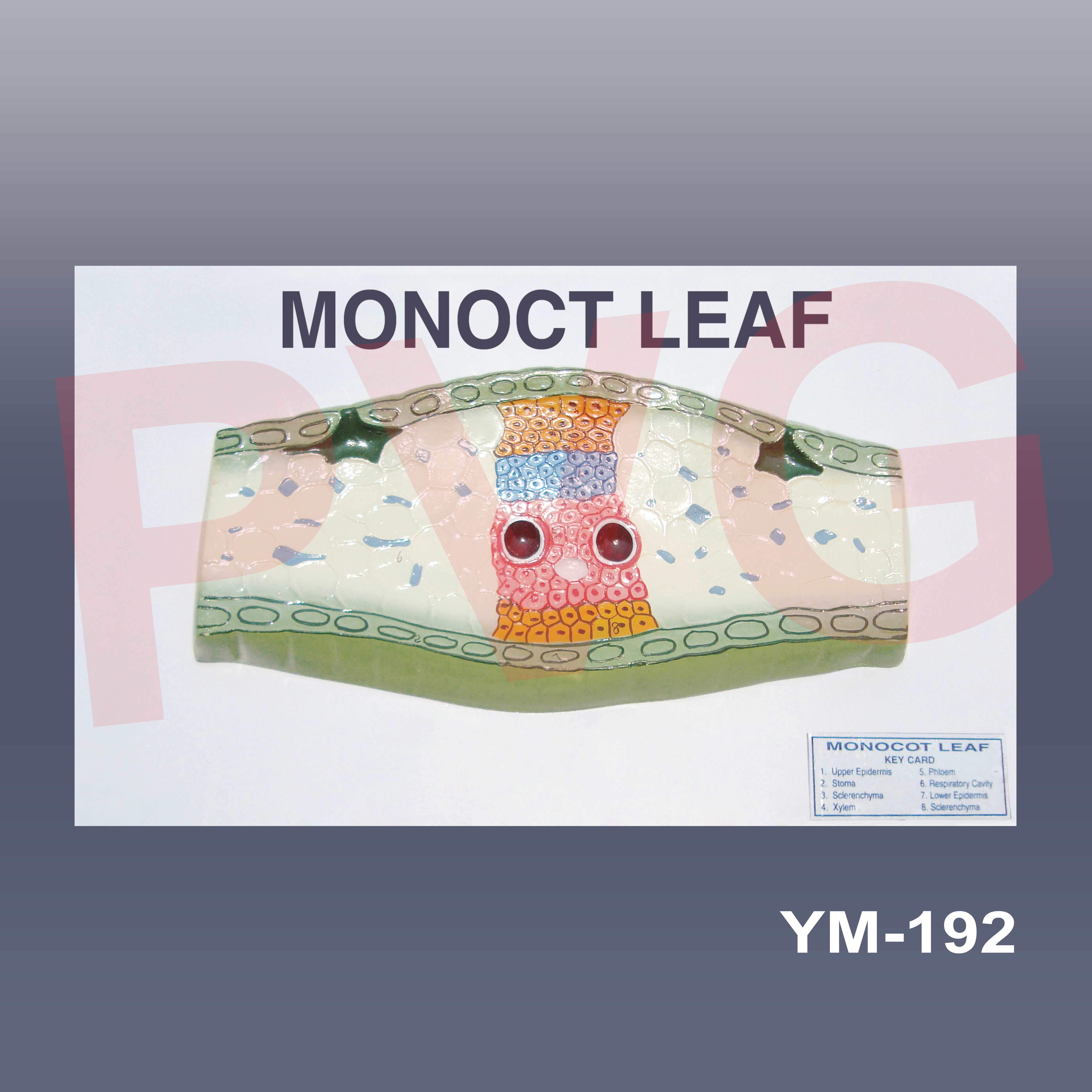
DICOT LEAF T.S
Transverse section Dicot Leaf Model with full Anatomical details of each layer.
Epidermis with stomata, Mesophyll layer i.e spongy parenchyma and palisade parenchyma and vascular Bundle.
The epidermis is shown on both the upper and lower surfaces of a leaf with thin cuticles which protect the plants against mechanical and physical injury.
In between the upper and lower epidermis is depicted a layer called the mesophyll. It is made up of parenchymatous cells and consists of chloroplasts that perform photosynthesis.
There are a number of spaces and cavities Shown in between these cells known as air cavities.
The vascular tissues (xylem and phloem) are shown in vascular bundles and are in the veins and midrib region.
The vascular bundles are shown protected by bundle sheath cells that are composed of one or more layers of parenchyma cells.
Very elaborate model for Botanical students to understand easily the structure of Leaf and functioning of various parts
Mounted on baseboard with Key.
Be the first to review “YM-191 LEAF TRANSVERSE SECTION DICOT (ECONOMICAL)”
Related products
Anatomical Models

Anatomical Models

Anatomical Models
Anatomical Models

Anatomical Models
Anatomical Models
Anatomical Models
Anatomical Models
Reviews
There are no reviews yet.