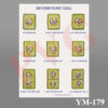
Skip to content
PVG INTERNATIONALPVG INTERNATIONAL
    • Mobile: +91-9466446612 +91-9728497962
    • E-Mail: info@pvginternationals.com info@narayanienterprises.com
  • Cart

    No products in the cart.

  • Home
  • About Us
  • Certifications
  • Products
  • Contact
  • Blog
Add to wishlist
MODEL OF PLANT CELL Enlarged Anatomical model as seen under the Electron Microscope mounted on base board with key card. All the Parts i.e Nucleus, Vacuoles Cell membrane, Endoplasmic Reticulum, Mitochondrion. Golgi apparatus, Chloroplast are detailed in this model and numbered too. Functions of all parts can be detailed easily by this Model e.g Chloroplast which makes plants green and is for Photosynthesis i.e take sunlight and use it to turn water and carbon dioxide into food through a process called photosynthesis. A plant cell is a tiny building block that makes up all parts of a plant, just like how a brick is used to build a house. Each plant is made up of millions of these cells, and each cell has special parts that help it do its job. Thick outer layer called the cell wall is shown, which gives them strength and helps them keep their shape. Inside the plant cell, is a  a jelly-like substance called cytoplasm, which holds all the important parts, or organelles, that help the cell work properly. This is how plants make their own food and release oxygen into the air, which is great for us to breathe! Another important part of the plant cell is Nucleus the centre point of any plant cellThe cell vacuole, which is like a big storage bag. It holds water, nutrients, and waste products. The vacuole is usually much larger in plant cells compared to animal cells, and it helps keep the plant firm and strong by maintaining pressure. All these parts work together to help the plant grow, heal, and produce flowers, fruits, and seeds. So, plant cells are really amazing little structures! They work hard every day to help the plant stay healthy and grow tall. Understanding how plant cells function helps us appreciate the important role that plants play in our world, from providing food to giving us clean air. Students when see any plant, can understand about all the small cells inside it working together to keep it alive!
Home / Anatomical Models

YM-177 3D MODEL OF PLANT CELL

  •  MODEL OF PLANT MITOSIS. Model depicts how a cell divides into two with 9 stages show the cell division of a plant ,mounted on board. Mitosis is process in the cell cycle where the newly formed DNA is separated and two new cells are formed with the same number and kind of chromosomes as in the parent nucleus. Mitosis is a process of asexual reproduction observed in unicellular organisms. Mitosis is essential for the growth of the cells and the replacement of worn-out cells. In plants, mitosis leads to the growth of vegetative parts of the plant like root tip, stem tip, etc. The different stages of mitosis occurring during  the cell division are Interphase,Prophase,Metaphase,Anaphase,Telophase,Cytokinesis,Daughter cells are shown. All phases are in 3D shape represented and cut to show the inside of the cell especially nucleus and Chromatin material. Movement of Chromosomes are easily seen from parent cell to Daughter cells Mitosis helps in Equal distribution of chromosomes to each daughter cell. It is responsible for the growth and development of the plant. It maintains the constant number of chromosomes in all the cells of a plant. Mitosis is required for asexual reproduction, vegetative propagation in plants and is also responsible for the repair and regeneration of damaged tissues. Mitosis helps in maintaining the purity of the genome as no recombination or crossing over takes place.
  • HUMAN EYE WITH ORBIT Versatile Model,Extremelyusefull,Dissectable model exhibiting complete Anatomy of Eye with bony orbit ,For Medical Students LIFE-LIKE OPTIC NERVE: Demonstrates  the optic nerve authentically within the bony orbit of the eye, capturing the natural position of the bony floor and medial wall. ENHANCED ANATOMICAL DEPICTIONS: Perfect for Teachers educators and medical professionals, to teach the students with our model brings anatomy to life, providing a tangible and visually engaging tool for demonstrations. 8 parts DISSECTIBLE MODEL: Dive deep into the intricacies of the eye's anatomy with our dissectible model, allowing for a comprehensive exploration of its various parts. FOUR TIMES ENLARGED MODEL: EnhancesStudents learning experience , enabling a detailed examination of the eye's structure for better understanding.

3 Dimensional, detailed as seen under an electron microscope
,mounted on board size 14 x 10 x 3 1/2” high, with key.

SKU: YM-177 PLANT CELL Category: Anatomical Models
  •  MODEL OF PLANT MITOSIS. Model depicts how a cell divides into two with 9 stages show the cell division of a plant ,mounted on board. Mitosis is process in the cell cycle where the newly formed DNA is separated and two new cells are formed with the same number and kind of chromosomes as in the parent nucleus. Mitosis is a process of asexual reproduction observed in unicellular organisms. Mitosis is essential for the growth of the cells and the replacement of worn-out cells. In plants, mitosis leads to the growth of vegetative parts of the plant like root tip, stem tip, etc. The different stages of mitosis occurring during  the cell division are Interphase,Prophase,Metaphase,Anaphase,Telophase,Cytokinesis,Daughter cells are shown. All phases are in 3D shape represented and cut to show the inside of the cell especially nucleus and Chromatin material. Movement of Chromosomes are easily seen from parent cell to Daughter cells Mitosis helps in Equal distribution of chromosomes to each daughter cell. It is responsible for the growth and development of the plant. It maintains the constant number of chromosomes in all the cells of a plant. Mitosis is required for asexual reproduction, vegetative propagation in plants and is also responsible for the repair and regeneration of damaged tissues. Mitosis helps in maintaining the purity of the genome as no recombination or crossing over takes place.
  • HUMAN EYE WITH ORBIT Versatile Model,Extremelyusefull,Dissectable model exhibiting complete Anatomy of Eye with bony orbit ,For Medical Students LIFE-LIKE OPTIC NERVE: Demonstrates  the optic nerve authentically within the bony orbit of the eye, capturing the natural position of the bony floor and medial wall. ENHANCED ANATOMICAL DEPICTIONS: Perfect for Teachers educators and medical professionals, to teach the students with our model brings anatomy to life, providing a tangible and visually engaging tool for demonstrations. 8 parts DISSECTIBLE MODEL: Dive deep into the intricacies of the eye's anatomy with our dissectible model, allowing for a comprehensive exploration of its various parts. FOUR TIMES ENLARGED MODEL: EnhancesStudents learning experience , enabling a detailed examination of the eye's structure for better understanding.
Categories
  • Chemistry - General & Chemical Laboratory Apparatus
  • Laboratory Glassware
    • Distillation Assembly & Rotary Evaporators
    • Laboratory Glassware section -1
    • Interchangeable Laboratory Glassware
    • Organic Chemistry Set
    • Clevenger, Dean & Stark
    • Chromatography Columns & Gas Analysis
    • Clinical Laboratoy Glassware
    • Dairy & Milk Testing
    • Hydrometers
    • Thermometers
    • Quartz Glassware
    • Sintered Glasswae
    • Reaction Vesssel
    • Volumetric Glasswares
  • Anatomical Models
  • Biological Apparatus & Material
  • Microscopes & Accessories
  • Physics & Electronic Laboratory Apparatus
  • Pharmacy Apparatus
  • Earth Sciences
  • Plasticware
  • Uncategories
  • Agriculture Equipment
  • Surgical Instruments & Hospital Equipment
  • Chemicals
  • Material Testing
Recently Viewed
  • Cat.No. Description 15.01 Syringe Filter Holder for 13 mm. membrane 15.02 Syringe Filter Holder for 25 mm. membrane 15.03 Vacuum Filter Holder for 47 mm. w/o Flask 15.04 3 Branch Sterility Test, for 47 mm. membrane 15.05 6 Branch Sterility Test, for 47 mm. membrane VACUUM FILTER HOLDERS, STAINLESS STEEL (TO HOLD MEMBRANE FILTERS)
  • Comprises a borosilicate glass U tube with 225 mm. height, with each limb graduated 0-120 x 1 mm. Bend fitted with a filling tube and stopcock. Two platinum electrodes 14 mm. diameter can be adjusted at any position by locking PM-315 PVG Y CONNECTOR GLASS, PLAIN/ RIFFL
  • 150 mm steel with wooden handle. Flat Stainless steel Spatula for mixing for Ointment Slab NPH-53 OINTMENT SPATULA
  • Description
  • Reviews (0)

MODEL OF PLANT CELL

Enlarged Anatomical model as seen under the Electron Microscope mounted on base board with key card.

All the Parts i.e Nucleus, Vacuoles Cell membrane, Endoplasmic Reticulum, Mitochondrion. Golgi apparatus, Chloroplast are detailed in this model and numbered too.

Functions of all parts can be detailed easily by this Model e.g Chloroplast which makes plants green and is for Photosynthesis i.e take sunlight and use it to turn water and carbon dioxide into food through a process called photosynthesis.

A plant cell is a tiny building block that makes up all parts of a plant, just like how a brick is used to build a house. Each plant is made up of millions of these cells, and each cell has special parts that help it do its job.

Thick outer layer called the cell wall is shown, which gives them strength and helps them keep their shape.

Inside the plant cell, is a  a jelly-like substance called cytoplasm, which holds all the important parts, or organelles, that help the cell work properly.

This is how plants make their own food and release oxygen into the air, which is great for us to breathe!

Another important part of the plant cell is Nucleus the centre point of any plant cellThe cell vacuole, which is like a big storage bag. It holds water, nutrients, and waste products. The vacuole is usually much larger in plant cells compared to animal cells, and it helps keep the plant firm and strong by maintaining pressure. All these parts work together to help the plant grow, heal, and produce flowers, fruits, and seeds.

So, plant cells are really amazing little structures! They work hard every day to help the plant stay healthy and grow tall. Understanding how plant cells function helps us appreciate the important role that plants play in our world, from providing food to giving us clean air.

Students when see any plant, can understand about all the small cells inside it working together to keep it alive!

Reviews

There are no reviews yet.

Be the first to review “YM-177 3D MODEL OF PLANT CELL” Cancel reply

Related products

YM-12 VERTEBRAL COLOUMN WITH PELVIS THIS MODEL SHOWS ALL SIGNIFICANT FEATURES OF EACH VERTEBRA, , A  HERNIATED DISC AND VERTEBRAL NOTCH ETC. SPECIAL FEATURES INCLUDE: FLEXIBLE 29" TALL VERTEBRAL COLUMN COMPLETE WITH PELVIS, SACRUM, OCCIPITAL BONE, HERNIATED LUMBAR  DISC. DELUXE CHROME STANDS 34" HIGH   YM-12 A VERTEBRAL COLOUMN WITH PELVIS THIS MODEL SHOWS ALL SIGNIFICANT FEATURES OF EACH VERTEBRA, INCLUDING SPINAL CORD, NERVE ROOTS, THE VERTEBRAL ARTERY, A  HERNIATED DISC AND VERTEBRAL NOTCH ETC. SPECIAL FEATURES INCLUDE: FLEXIBLE 29" TALL VERTEBRAL COLUMN COMPLETE WITH PELVIS, SACRUM, OCCIPITAL BONE, VERTEBRAL ARTERY, ALL NERVE BRANCHES AND HERNIATED LUMBAR DISC. DELUXE CHROME STANDS 34" HIGH
Add to wishlist
Quick View

Anatomical Models

YM-12 HUMAN VERTEBRAL COLUMN

Set of two, left and right feet bones, on base.
Add to wishlist
Quick View

Anatomical Models

YM-13 HUMAN FEET BONES

Excellent Educational model to teach Medical students the various parts of skull, its anatomy.Very helpful for Biology students to Understand the Anatomy of Skull. Superior model,three part, fibre glass, with full dentition and all other details. The calvaria is uncut and the lower jaw is spring held.
Add to wishlist
Quick View

Anatomical Models

YM-09 HUMAN SKULL

MODEL OF ANIMAL MITOSIS SET
Add to wishlist
Quick View

Anatomical Models

YM-30 ANIMAL MITOSIS SET

YM-26 CAT SKULL MODEL
Add to wishlist
Quick View

Anatomical Models

YM-26 CAT SKULL MODEL

Set of nine stages from blastoderm to yolk sac and stage shortly after hatching. On base size 12 x 20”.
Add to wishlist
Quick View

Anatomical Models

YM-42 LIFE CYCLE OF FISH

This model shown the osteoporosis and blood vessel.This model shown the osteoporosis and blood vessel.
Add to wishlist
Quick View

Anatomical Models

YM-20 FEMUR BONE

MODEL OF TYPICAL ANIMAL CELL
Add to wishlist
Quick View

Anatomical Models

YM-28 ANIMAL CELL

  • Home
  • About Us
  • Certifications
  • Products
  • Contact
  • Blog

Address Details

PVG International
# 6 - C, New Ram Nagar, Ambala Cantt - 133 001, Haryana, India.
# P.O. Box-113

Our Visitor

001079

Contact Details

  • info@pvginternationals.com
  • info@narayanienterprises.com
  • +91-9466446612
  • +91-9728497962
  • Phone: +91-171-2651006, 2653272
  • Fax: +91-171-2653259
Copyright 2025 © PVG INTERNATIONAL
  • Home
  • About Us
  • Certifications
  • Products
  • Contact
  • Blog
  • Login
    • Mobile: +91-9466446612 +91-9728497962
    • E-Mail: info@pvginternationals.com info@narayanienterprises.com

Login

captcha

Lost your password?

Register

Your personal data will be used to support your experience throughout this website, to manage access to your account, and for other purposes described in our privacy policy.

captcha