Skip to content
PVG INTERNATIONALPVG INTERNATIONAL
    • Mobile: +91-9466446612 +91-9728497962
    • E-Mail: info@pvginternationals.com info@narayanienterprises.com
  • Cart

    No products in the cart.

  • Home
  • About Us
  • Certifications
  • Products
  • Contact
  • Blog
Add to wishlist
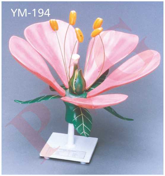
DICOT FLOWER MODEL Flowers represent an important adaptation for plants, as the different shapes and colors are used to attract different pollinating species of insects and other animals. The colorful flowers characteristic of dicots consist of a specific number of sepals, or petals. In dicots, the number of petals occurs in groups of four or five . PVG’s Typical Dicot Flower Model is enlarged, Educational, 3 D plastic, unbreakable model, this model is for  students to easily study the floral components and the reproductive functions in flowers. Model can be disassembled to reveal the pollen grains inside, showing the process of angiosperm pollination and fertilization . Important structures are numbered on the flower model and corresponds to an identification key. THIS Model includes structures of Flower like receptacle, petal, sepal, gynoecium (stigma and style), male stamen (anther and filament), pollen, pollen tube etc. Petals, sepals and  stamens are detachable. Ovary is dissected.  Hand painted each part in different color, to deepen the understanding of the structure of the dicotyledonous flower. Sepals are colored green, petals, pink, ovary green, and anthers with pollen tubes in yellow color. This Model is Botanical teaching aid for Dicot plants. Saves learning time and improve learning efficiency This is a perfect model for teachers to make students to understand the Anatomy of Flower in Botanical classes.    
Home / Anatomical Models

YM-194 DICOT FLOWER MODEL

  • Shows basic structure of a flower i.e. petal, sepal, stem, pistil, stamen and anther. Size 16 x 12”.

Highly enlarged, complete flower. Shows sepals, petals, stamens,
pistil, anther and dissected ovary. Petals, sepals and stamen are
detachable. Overall size 14 x 14 x 15”.

SKU: YM-194 DICOT FLOWER MODEL Category: Anatomical Models
  • Shows basic structure of a flower i.e. petal, sepal, stem, pistil, stamen and anther. Size 16 x 12”.
Categories
  • Chemistry - General & Chemical Laboratory Apparatus
  • Laboratory Glassware
    • Distillation Assembly & Rotary Evaporators
    • Laboratory Glassware section -1
    • Interchangeable Laboratory Glassware
    • Organic Chemistry Set
    • Clevenger, Dean & Stark
    • Chromatography Columns & Gas Analysis
    • Clinical Laboratoy Glassware
    • Dairy & Milk Testing
    • Hydrometers
    • Thermometers
    • Quartz Glassware
    • Sintered Glasswae
    • Reaction Vesssel
    • Volumetric Glasswares
  • Anatomical Models
  • Biological Apparatus & Material
  • Microscopes & Accessories
  • Physics & Electronic Laboratory Apparatus
  • Pharmacy Apparatus
  • Earth Sciences
  • Plasticware
  • Uncategories
  • Agriculture Equipment
  • Surgical Instruments & Hospital Equipment
  • Chemicals
  • Material Testing
Recently Viewed
  • NPH-08 ball mill motor driven It works on the principle of impact and attrition. Impact occurs when the balls are lifted by the rotating mill and then drop onto the material to be ground. The force of the impact breaks the material into smaller pieces. Attrition occurs when the balls rub against each other and against the material to be ground. Ball mills are commonly used for crushing and grinding the materials into an extremely fine form. The ball mill contains a hollow cylindrical shell that rotates about its axis. Robust, strong construction,For mixing & grinding, electrically operated. Instrument Powder coated with Heavy stable base,Fitted with Fractional Horse Power Motor Gear. With  Stainless steel jar.Capacity one kg and two Kg. Jar is interchangeable and easily removable and is light in weight . Compact unit to operate on 220 volts, 50 Hz A.C mains. NPH-08 TELE THERMOMETER
  • The instrument is designed to test two batches of six tablets, simultaneously. The unit is extremely useful for pharmaceutical industries. Extremely useful for pharmaceutical labs   It is used in quality control and R & D to determine compliance with the disintegration requirement of the tablet and capsule as per required standards. Instrument uses the latest microprocessor technology and advanced engineering techniques so as to give enhanced accuracy and reproducibility.   Microprocessor based, easy to use,With long trouble free life. Seven segment LED display Supports Two baskets assemblies. Temperature is controlled digitally so produces accurate results. The Tablet Disintegration Double Test Apparatus with a capacity of 1000 ml is usedfor testing the disintegration properties of tablets. Allows for simultaneous testing of multiple tablets, offering a high capacity of 1000ml for conducting disintegration tests. The apparatus consists of multiple test stations or Two baskets that can hold tablets during the disintegration process. The apparatus features a water bath or reservoir with temperature control to maintain the desired testing conditions. Measures Disintegration time of 12 tablets at one time. Disintegration time is displayed on Seven Segment LED display. Electrically operated,Robust powder coated Mild steel body.Front panel provided with all main controls.Supplied complete with all accessories and working manual. NPH-26 DISINTEGRATION TEST APPARATUS (DOUBLE BASKET )
  • Description
  • Reviews (0)

DICOT FLOWER MODEL

Flowers represent an important adaptation for plants, as the different shapes and colors are used to attract different pollinating species of insects and other animals. The colorful flowers characteristic of dicots consist of a specific number of sepals, or petals. In dicots, the number of petals occurs in groups of four or five .

PVG’s Typical Dicot Flower Model is enlarged, Educational, 3 D plastic, unbreakable model, this model is for  students to easily study the floral components and the reproductive functions in flowers.

Model can be disassembled to reveal the pollen grains inside, showing the process of angiosperm pollination and fertilization .

Important structures are numbered on the flower model and corresponds to an identification key.

THIS Model includes structures of Flower like receptacle, petal, sepal, gynoecium (stigma and style), male stamen (anther and filament), pollen, pollen tube etc.

Petals, sepals and  stamens are detachable. Ovary is dissected.  Hand painted each part in different color, to deepen the understanding of the structure of the dicotyledonous flower.

Sepals are colored green, petals, pink, ovary green, and anthers with pollen tubes in yellow color.

This Model is Botanical teaching aid for Dicot plants. Saves learning time and improve learning efficiency

This is a perfect model for teachers to make students to understand the Anatomy of Flower in Botanical classes.


Reviews

There are no reviews yet.

Be the first to review “YM-194 DICOT FLOWER MODEL” Cancel reply

Related products

KNEE FUNCTIONAL JOINT LIFE SIZE WITH MUSCLES
Add to wishlist
Quick View

Anatomical Models

YM-24 KNEE ANATOMY LIFE SIZE MODEL

Set of two, left and right hand bones, on base.
Add to wishlist
Quick View

Anatomical Models

YM-14 HUMAN HAND BONES

YM-12 VERTEBRAL COLOUMN WITH PELVIS THIS MODEL SHOWS ALL SIGNIFICANT FEATURES OF EACH VERTEBRA, , A  HERNIATED DISC AND VERTEBRAL NOTCH ETC. SPECIAL FEATURES INCLUDE: FLEXIBLE 29" TALL VERTEBRAL COLUMN COMPLETE WITH PELVIS, SACRUM, OCCIPITAL BONE, HERNIATED LUMBAR  DISC. DELUXE CHROME STANDS 34" HIGH   YM-12 A VERTEBRAL COLOUMN WITH PELVIS THIS MODEL SHOWS ALL SIGNIFICANT FEATURES OF EACH VERTEBRA, INCLUDING SPINAL CORD, NERVE ROOTS, THE VERTEBRAL ARTERY, A  HERNIATED DISC AND VERTEBRAL NOTCH ETC. SPECIAL FEATURES INCLUDE: FLEXIBLE 29" TALL VERTEBRAL COLUMN COMPLETE WITH PELVIS, SACRUM, OCCIPITAL BONE, VERTEBRAL ARTERY, ALL NERVE BRANCHES AND HERNIATED LUMBAR DISC. DELUXE CHROME STANDS 34" HIGH
Add to wishlist
Quick View

Anatomical Models

YM-12 HUMAN VERTEBRAL COLUMN

MODEL OF ANIMAL MITOSIS SET
Add to wishlist
Quick View

Anatomical Models

YM-30 ANIMAL MITOSIS SET

Fibre glass, depicting full details..
Add to wishlist
Quick View

Anatomical Models

YM- 10 HUMAN FETAL SKULL

Set of two, left and right feet bones, on base.
Add to wishlist
Quick View

Anatomical Models

YM-13 HUMAN FEET BONES

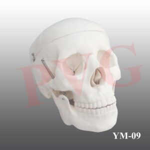
Excellent Educational model to teach Medical students the various parts of skull, its anatomy.Very helpful for Biology students to Understand the Anatomy of Skull. Superior model,three part, fibre glass, with full dentition and all other details. The calvaria is uncut and the lower jaw is spring held.
Add to wishlist
Quick View

Anatomical Models

YM-09 HUMAN SKULL

This model shown the osteoporosis and blood vessel.This model shown the osteoporosis and blood vessel.
Add to wishlist
Quick View

Anatomical Models

YM-20 FEMUR BONE

  • Home
  • About Us
  • Certifications
  • Products
  • Contact
  • Blog

Address Details

PVG International
# 6 - C, New Ram Nagar, Ambala Cantt - 133 001, Haryana, India.
# P.O. Box-113

Our Visitor

080773

Contact Details

  • info@pvginternationals.com
  • info@narayanienterprises.com
  • +91-9466446612
  • +91-9728497962
  • Phone: +91-171-2651006, 2653272
  • Fax: +91-171-2653259
Copyright 2026 © PVG INTERNATIONAL
  • Home
  • About Us
  • Certifications
  • Products
  • Contact
  • Blog
  • Login
    • Mobile: +91-9466446612 +91-9728497962
    • E-Mail: info@pvginternationals.com info@narayanienterprises.com

Login

captcha

Lost your password?

Register

Your personal data will be used to support your experience throughout this website, to manage access to your account, and for other purposes described in our privacy policy.

captcha