
YM-208 DNA STRUCTURE MODEL


Simple, easy to understand double helix model. A three dimensional model with color coded plastic pieces showing base
pairing i.e. nitrogenous bases (Adenine, Thiamine, Guanine,
Cytosine), and pairs attached representing hydrogen bonds and
phosphates. On stand approx. 350 mm high.
MODEL OF DNA
This models resembles the structure of DNA, as represented in Watson and Crick’s model, is a double-stranded, antiparallel, right-handed helix.
Double Helix Colorfull , 3 D ,Plastic model,very usefull model and easy to explain the students.
- Color-coded components to differentiate between the different elements of DNA, aiding in understanding.
- Nitrogenous base pairing in correct sequence is clearly depicted, attached by Hydrogen bonds and phosphates
- Two flexible strands signifying alternating pentose and phosphate units attach to the end of each pair, forming the double helix.
Be the first to review “YM-208 DNA STRUCTURE MODEL”
Related products

Anatomical Models
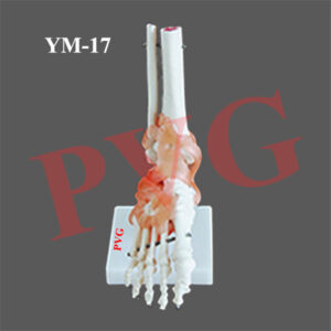
Anatomical Models
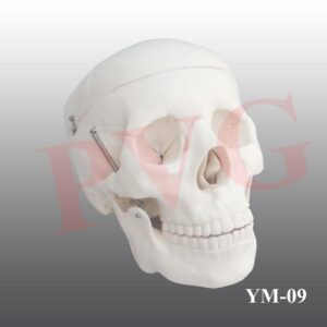
Anatomical Models

Anatomical Models

Anatomical Models
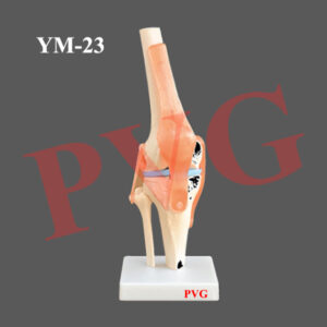
Anatomical Models

Anatomical Models

Anatomical Models
Reviews
There are no reviews yet.