
YM-165 MODEL OF GIANT EYE
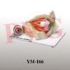

Enlarged 6 times, the different part of the eyeball model are
detachable to show the Tunica external: showing cornea and sclera
with attachments of ocular muscles and optic nerve; Tunica media:
showing the iris, the ciliary body and the choroid; Tunica internal is
retina; Refraction media: Showing the leans the vitreous body.
Mounted on stand
Be the first to review “YM-165 MODEL OF GIANT EYE”
Related products

Anatomical Models
YM-04 HUMAN SKELETON WITH NERVES & BLOOD VESSELS (TALL 85 cms)
Anatomical Models

Anatomical Models
Anatomical Models

Anatomical Models
Anatomical Models

Anatomical Models
Anatomical Models
Reviews
There are no reviews yet.