Skip to content
PVG INTERNATIONALPVG INTERNATIONAL
    • Mobile: +91-9466446612 +91-9728497962
    • E-Mail: info@pvginternationals.com info@narayanienterprises.com
  • Cart

    No products in the cart.

  • Home
  • About Us
  • Certifications
  • Products
  • Contact
  • Blog
Add to wishlist
Awaiting product image
Home / Anatomical Models

YM-183 ROOT, STEM AND LEAF

  • MODEL OF DICOT STEM The dicot stem model in Transverse and Longitudinal section, is a vibrantly colored 3D model with detail that shows the inner anatomy and vasculature of a dicot stem Teaching model Showing all details of cellular structures, a very useful and instructive model for the study of  students in Botany classes All the parts i.e Epidermis, cuticle, Cortex, Endodermis, Pericycle, Phloem, Cambium Metaxylem, Protoxylem and Pith are numbered and Model is mounted on a base board for display with key card
  • The life cycle of Penicillium

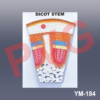
Shows root penetrating soil and stem partly dissected. Sectioned
leaf reveals epidermis, palisades and spongy cover. Size 18 x 25”.

SKU: YM-183 ROOT, STEM AND LEAF Category: Anatomical Models
  • MODEL OF DICOT STEM The dicot stem model in Transverse and Longitudinal section, is a vibrantly colored 3D model with detail that shows the inner anatomy and vasculature of a dicot stem Teaching model Showing all details of cellular structures, a very useful and instructive model for the study of  students in Botany classes All the parts i.e Epidermis, cuticle, Cortex, Endodermis, Pericycle, Phloem, Cambium Metaxylem, Protoxylem and Pith are numbered and Model is mounted on a base board for display with key card
  • The life cycle of Penicillium
Categories
  • Chemistry - General & Chemical Laboratory Apparatus
  • Laboratory Glassware
    • Distillation Assembly & Rotary Evaporators
    • Laboratory Glassware section -1
    • Interchangeable Laboratory Glassware
    • Organic Chemistry Set
    • Clevenger, Dean & Stark
    • Chromatography Columns & Gas Analysis
    • Clinical Laboratoy Glassware
    • Dairy & Milk Testing
    • Hydrometers
    • Thermometers
    • Quartz Glassware
    • Sintered Glasswae
    • Reaction Vesssel
    • Volumetric Glasswares
  • Anatomical Models
  • Biological Apparatus & Material
  • Microscopes & Accessories
  • Physics & Electronic Laboratory Apparatus
  • Pharmacy Apparatus
  • Earth Sciences
  • Plasticware
  • Uncategories
  • Agriculture Equipment
  • Surgical Instruments & Hospital Equipment
  • Chemicals
  • Material Testing
Recently Viewed
  • Placeholder YM-183 ROOT, STEM AND LEAF
  • MODEL OF PLANT CELL Enlarged Anatomical model as seen under the Electron Microscope mounted on base board with key card. All the Parts i.e Nucleus, Vacuoles Cell membrane, Endoplasmic Reticulum, Mitochondrion. Golgi apparatus, Chloroplast are detailed in this model and numbered too. Functions of all parts can be detailed easily by this Model e.g Chloroplast which makes plants green and is for Photosynthesis i.e take sunlight and use it to turn water and carbon dioxide into food through a process called photosynthesis. A plant cell is a tiny building block that makes up all parts of a plant, just like how a brick is used to build a house. Each plant is made up of millions of these cells, and each cell has special parts that help it do its job. Thick outer layer called the cell wall is shown, which gives them strength and helps them keep their shape. Inside the plant cell, is a  a jelly-like substance called cytoplasm, which holds all the important parts, or organelles, that help the cell work properly. This is how plants make their own food and release oxygen into the air, which is great for us to breathe! Another important part of the plant cell is Nucleus the centre point of any plant cellThe cell vacuole, which is like a big storage bag. It holds water, nutrients, and waste products. The vacuole is usually much larger in plant cells compared to animal cells, and it helps keep the plant firm and strong by maintaining pressure. All these parts work together to help the plant grow, heal, and produce flowers, fruits, and seeds. So, plant cells are really amazing little structures! They work hard every day to help the plant stay healthy and grow tall. Understanding how plant cells function helps us appreciate the important role that plants play in our world, from providing food to giving us clean air. Students when see any plant, can understand about all the small cells inside it working together to keep it alive! YM-177 3D MODEL OF PLANT CELL
  • Reviews (0)

Reviews

There are no reviews yet.

Be the first to review “YM-183 ROOT, STEM AND LEAF” Cancel reply

Related products

YM- 41 FISH MODEL
Add to wishlist
Quick View

Anatomical Models

YM- 41 FISH MODEL

Miniature skeleton model offer unique hands-on learning experience for young scientists, curious kids or medical professionals. Include a movable jaw on springs, a skull whose top half opens for a look inside, arms and legs that are removable for individual study. Mounted on stand. Made of washable and unbreakable PVC plastic.
Add to wishlist
Quick View

Anatomical Models

YM- 06 MODEL OF MINI HUMAN SKELETON

Fibre glass, complete with mandible on base, with illustrated key.
Add to wishlist
Quick View

Anatomical Models

YM-25 GOAT SKULL MODEL

YM-12 VERTEBRAL COLOUMN WITH PELVIS THIS MODEL SHOWS ALL SIGNIFICANT FEATURES OF EACH VERTEBRA, , A  HERNIATED DISC AND VERTEBRAL NOTCH ETC. SPECIAL FEATURES INCLUDE: FLEXIBLE 29" TALL VERTEBRAL COLUMN COMPLETE WITH PELVIS, SACRUM, OCCIPITAL BONE, HERNIATED LUMBAR  DISC. DELUXE CHROME STANDS 34" HIGH   YM-12 A VERTEBRAL COLOUMN WITH PELVIS THIS MODEL SHOWS ALL SIGNIFICANT FEATURES OF EACH VERTEBRA, INCLUDING SPINAL CORD, NERVE ROOTS, THE VERTEBRAL ARTERY, A  HERNIATED DISC AND VERTEBRAL NOTCH ETC. SPECIAL FEATURES INCLUDE: FLEXIBLE 29" TALL VERTEBRAL COLUMN COMPLETE WITH PELVIS, SACRUM, OCCIPITAL BONE, VERTEBRAL ARTERY, ALL NERVE BRANCHES AND HERNIATED LUMBAR DISC. DELUXE CHROME STANDS 34" HIGH
Add to wishlist
Quick View

Anatomical Models

YM-12 HUMAN VERTEBRAL COLUMN

YM-18 HUMAN KNEE JOINT
Add to wishlist
Quick View

Anatomical Models

YM-18 HUMAN KNEE JOINT

MODEL OF TYPICAL ANIMAL CELL
Add to wishlist
Quick View

Anatomical Models

YM-28 ANIMAL CELL

Cross section mounted on base height approx. 11”, with key.
Add to wishlist
Quick View

Anatomical Models

YM-29 ANIMAL CELL MODEL

Excellent Educational model to teach Medical students the various parts of skull, its anatomy.Very helpful for Biology students to Understand the Anatomy of Skull. Superior model,three part, fibre glass, with full dentition and all other details. The calvaria is uncut and the lower jaw is spring held.
Add to wishlist
Quick View

Anatomical Models

YM-09 HUMAN SKULL

  • Home
  • About Us
  • Certifications
  • Products
  • Contact
  • Blog

Address Details

PVG International
# 6 - C, New Ram Nagar, Ambala Cantt - 133 001, Haryana, India.
# P.O. Box-113

Our Visitor

016286

Contact Details

  • info@pvginternationals.com
  • info@narayanienterprises.com
  • +91-9466446612
  • +91-9728497962
  • Phone: +91-171-2651006, 2653272
  • Fax: +91-171-2653259
Copyright 2026 © PVG INTERNATIONAL
  • Home
  • About Us
  • Certifications
  • Products
  • Contact
  • Blog
  • Login
    • Mobile: +91-9466446612 +91-9728497962
    • E-Mail: info@pvginternationals.com info@narayanienterprises.com

Login

captcha

Lost your password?

Register

Your personal data will be used to support your experience throughout this website, to manage access to your account, and for other purposes described in our privacy policy.

captcha