Skip to content
PVG INTERNATIONALPVG INTERNATIONAL
    • Mobile: +91-9466446612 +91-9728497962
    • E-Mail: info@pvginternationals.com info@narayanienterprises.com
  • Cart

    No products in the cart.

  • Home
  • About Us
  • Certifications
  • Products
  • Contact
  • Blog
Add to wishlist
MODEL OF PARAMECIUM Paramecium are complex single-celled organisms that live in ponds that predate bacteria and smaller unicellular organisms. They are covered with cilia .used for for movement and use a mouth-like oral groove to catch their prey, breaking it down and expelling the waste. Educational Protozoan ciliated model well suited for Biology class students With clear depiction of internal as well as external structure. Fibreglass, Multicolored Model mounted on base board with detailed design. This paramecium protozoa is a high quality model that will enhance details and realism to any of the rendering projects. In this Model all the parts i.e Cell membrane, Contracting Vacuole, cilia,Mitochondria, nucleus,Oral groove, Food vacuole are clearly depicted and colored for easy understanding by Students.
Home / Anatomical Models

YM-35 PARAMECIUM

  • Three models on a 18 x 12” base showing the anatomy of Hydra in details, with manual.
  • MODEL OF AMOEBA Excellent colored display model for kids in classrooms, school colleges on base board Simplified representation of the microscopic single-celled organism for educational or research purposes. Model is Economical, Light weight and easy to carry even in fields to educate general public for Amoebic diseases. Clearly depicts the internal structure of this Unicellular Amoeba with painted different colors.MODEL OF AMOEBA Excellent colored display model for kids in classrooms, school colleges on base board Simplified representation of the microscopic single-celled organism for educational or research purposes. Model is Economical, Light weight and easy to carry even in fields to educate general public for Amoebic diseases. Clearly depicts the internal structure of this Unicellular Amoeba with painted different colors.

Depicts all the basic feature. Mounted on a 10 x 15” base, with
manual.

SKU: YM-35 PARAMECIUM Category: Anatomical Models
  • Three models on a 18 x 12” base showing the anatomy of Hydra in details, with manual.
  • MODEL OF AMOEBA Excellent colored display model for kids in classrooms, school colleges on base board Simplified representation of the microscopic single-celled organism for educational or research purposes. Model is Economical, Light weight and easy to carry even in fields to educate general public for Amoebic diseases. Clearly depicts the internal structure of this Unicellular Amoeba with painted different colors.MODEL OF AMOEBA Excellent colored display model for kids in classrooms, school colleges on base board Simplified representation of the microscopic single-celled organism for educational or research purposes. Model is Economical, Light weight and easy to carry even in fields to educate general public for Amoebic diseases. Clearly depicts the internal structure of this Unicellular Amoeba with painted different colors.
Categories
  • Chemistry - General & Chemical Laboratory Apparatus
  • Laboratory Glassware
    • Distillation Assembly & Rotary Evaporators
    • Laboratory Glassware section -1
    • Interchangeable Laboratory Glassware
    • Organic Chemistry Set
    • Clevenger, Dean & Stark
    • Chromatography Columns & Gas Analysis
    • Clinical Laboratoy Glassware
    • Dairy & Milk Testing
    • Hydrometers
    • Thermometers
    • Quartz Glassware
    • Sintered Glasswae
    • Reaction Vesssel
    • Volumetric Glasswares
  • Anatomical Models
  • Biological Apparatus & Material
  • Microscopes & Accessories
  • Physics & Electronic Laboratory Apparatus
  • Pharmacy Apparatus
  • Earth Sciences
  • Plasticware
  • Uncategories
  • Agriculture Equipment
  • Surgical Instruments & Hospital Equipment
  • Chemicals
  • Material Testing
Recently Viewed
  • MODEL OF PARAMECIUM Paramecium are complex single-celled organisms that live in ponds that predate bacteria and smaller unicellular organisms. They are covered with cilia .used for for movement and use a mouth-like oral groove to catch their prey, breaking it down and expelling the waste. Educational Protozoan ciliated model well suited for Biology class students With clear depiction of internal as well as external structure. Fibreglass, Multicolored Model mounted on base board with detailed design. This paramecium protozoa is a high quality model that will enhance details and realism to any of the rendering projects. In this Model all the parts i.e Cell membrane, Contracting Vacuole, cilia,Mitochondria, nucleus,Oral groove, Food vacuole are clearly depicted and colored for easy understanding by Students. YM-35 PARAMECIUM
  • An open model with all parts readily observable of d.c electrical motor, an armature wound with enamelled copper wire and permanent magnetic field by a removable bar magnet, external connections by a pair of 4mm. socket terminals. Operates on 4-6 v D.C. Overall height 180mm. MS-469 PVG DEMONSTRATION ELECTRIC MOTOR
  • Description
  • Reviews (0)

MODEL OF PARAMECIUM

Paramecium are complex single-celled organisms that live in ponds that predate bacteria and smaller unicellular organisms. They are covered with cilia .used for for movement and use a mouth-like oral groove to catch their prey, breaking it down and expelling the waste.

Educational Protozoan ciliated model well suited for Biology class students

With clear depiction of internal as well as external structure.

Fibreglass, Multicolored Model mounted on base board with detailed design.

This paramecium protozoa is a high quality model that will enhance details and realism to any of the rendering projects.

In this Model all the parts i.e Cell membrane, Contracting Vacuole, cilia,Mitochondria, nucleus,Oral groove, Food vacuole are clearly depicted and colored for easy understanding by Students.

Reviews

There are no reviews yet.

Be the first to review “YM-35 PARAMECIUM” Cancel reply

Related products

Set of nine stages from blastoderm to yolk sac and stage shortly after hatching. On base size 12 x 20”.
Add to wishlist
Quick View

Anatomical Models

YM-42 LIFE CYCLE OF FISH

Fibre glass, complete with mandible on base, with illustrated key.
Add to wishlist
Quick View

Anatomical Models

YM-25 GOAT SKULL MODEL

YM- 41 FISH MODEL
Add to wishlist
Quick View

Anatomical Models

YM- 41 FISH MODEL

Placeholder
Add to wishlist
Quick View

Anatomical Models

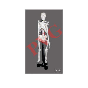
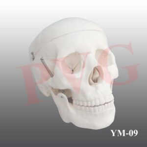
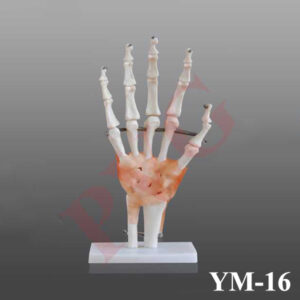
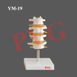
YM-04 HUMAN SKELETON WITH NERVES & BLOOD VESSELS (TALL 85 cms)

ELBOW JOINT LIFE SIZE WITH MUSCLES
Add to wishlist
Quick View

Anatomical Models

YM-22 ELBOW JOINT LIFE SIZE WITH MUSCLES

Set of two, left and right feet bones, on base.
Add to wishlist
Quick View

Anatomical Models

YM-13 HUMAN FEET BONES

YM-26 CAT SKULL MODEL
Add to wishlist
Quick View

Anatomical Models

YM-26 CAT SKULL MODEL

KNEE JOINT LIFE SIZE WITH MUSCLES
Add to wishlist
Quick View

Anatomical Models

YM-23 KNEE JOINT LIFE SIZE WITH MUSCLES

  • Home
  • About Us
  • Certifications
  • Products
  • Contact
  • Blog

Address Details

PVG International
# 6 - C, New Ram Nagar, Ambala Cantt - 133 001, Haryana, India.
# P.O. Box-113

Our Visitor

001069

Contact Details

  • info@pvginternationals.com
  • info@narayanienterprises.com
  • +91-9466446612
  • +91-9728497962
  • Phone: +91-171-2651006, 2653272
  • Fax: +91-171-2653259
Copyright 2025 © PVG INTERNATIONAL
  • Home
  • About Us
  • Certifications
  • Products
  • Contact
  • Blog
  • Login
    • Mobile: +91-9466446612 +91-9728497962
    • E-Mail: info@pvginternationals.com info@narayanienterprises.com

Login

captcha

Lost your password?

Register

Your personal data will be used to support your experience throughout this website, to manage access to your account, and for other purposes described in our privacy policy.

captcha