
YM-198 MODEL OF DICOT AND MONOCOT ROOT

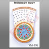
Transverse sectioned models of both mounted on one base board
for easy comparison of students. Size 12 x 24 x 3”.
Be the first to review “YM-198 MODEL OF DICOT AND MONOCOT ROOT”
Related products
Anatomical Models
Anatomical Models

Anatomical Models
Anatomical Models

Anatomical Models
Anatomical Models

Anatomical Models

Anatomical Models
Reviews
There are no reviews yet.