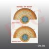
Skip to content
PVG INTERNATIONALPVG INTERNATIONAL
    • Mobile: +91-9466446612 +91-9728497962
    • E-Mail: info@pvginternationals.com info@narayanienterprises.com
  • Cart

    No products in the cart.

  • Home
  • About Us
  • Certifications
  • Products
  • Contact
  • Blog
Add to wishlist
MONOCOT ROOT Transverse section Colored Model Mounted on base board to show the full details of Monocot Root Anatomy.   This Model shows Structures like  Epiblema, Cortex, Endodermis, Pericycle, Pith, Vascular bundles. Xylem and Phloem are shown.   In This PVG’s Model of monocot root, the vascular bundles are arranged in a circular pattern   Vascular tissue is made up of alternate strands of xylem and phloem.   Vascular bundles are radial type shown in this Model.   Both Protoxylem and Metaxylem are shown.   Students can differentiate easily between Dicot and Monocot Root with these Models
Home / Anatomical Models

YM-197 MODEL OF MONOCOT ROOT

  • Transverse sectioned models of both mounted on one base board for easy comparison of students. Size 12 x 24 x 3”.
  • Model of dicot root

Transverse sectioned model, depicting cells in cross section.
Mounted on wooden base board. Size 10 x 12”.

SKU: YM-197 MONOCOT ROOT Category: Anatomical Models
  • Transverse sectioned models of both mounted on one base board for easy comparison of students. Size 12 x 24 x 3”.
  • Model of dicot root
Categories
  • Chemistry - General & Chemical Laboratory Apparatus
  • Laboratory Glassware
    • Distillation Assembly & Rotary Evaporators
    • Laboratory Glassware section -1
    • Interchangeable Laboratory Glassware
    • Organic Chemistry Set
    • Clevenger, Dean & Stark
    • Chromatography Columns & Gas Analysis
    • Clinical Laboratoy Glassware
    • Dairy & Milk Testing
    • Hydrometers
    • Thermometers
    • Quartz Glassware
    • Sintered Glasswae
    • Reaction Vesssel
    • Volumetric Glasswares
  • Anatomical Models
  • Biological Apparatus & Material
  • Microscopes & Accessories
  • Physics & Electronic Laboratory Apparatus
  • Pharmacy Apparatus
  • Earth Sciences
  • Plasticware
  • Uncategories
  • Agriculture Equipment
  • Surgical Instruments & Hospital Equipment
  • Chemicals
  • Material Testing
  • Description
  • Reviews (0)

MONOCOT ROOT

Transverse section Colored Model Mounted on base board to show the full details of Monocot Root Anatomy.


This Model shows Structures like  Epiblema, Cortex, Endodermis, Pericycle, Pith, Vascular bundles.

Xylem and Phloem are shown.


In This PVG’s Model of monocot root, the vascular bundles are arranged in a circular pattern


Vascular tissue is made up of alternate strands of xylem and phloem.


Vascular bundles are radial type shown in this Model.


Both Protoxylem and Metaxylem are shown.


Students can differentiate easily between Dicot and Monocot Root with these Models

Reviews

There are no reviews yet.

Be the first to review “YM-197 MODEL OF MONOCOT ROOT” Cancel reply

Related products

YM- 41 FISH MODEL
Add to wishlist
Quick View

Anatomical Models

YM- 41 FISH MODEL

Excellent Educational model to teach Medical students the various parts of skull, its anatomy.Very helpful for Biology students to Understand the Anatomy of Skull. Superior model,three part, fibre glass, with full dentition and all other details. The calvaria is uncut and the lower jaw is spring held.
Add to wishlist
Quick View

Anatomical Models

YM-09 HUMAN SKULL

This model shown the osteoporosis and blood vessel.This model shown the osteoporosis and blood vessel.
Add to wishlist
Quick View

Anatomical Models

YM-20 FEMUR BONE

Set of nine stages from blastoderm to yolk sac and stage shortly after hatching. On base size 12 x 20”.
Add to wishlist
Quick View

Anatomical Models

YM-42 LIFE CYCLE OF FISH

KNEE FUNCTIONAL JOINT LIFE SIZE WITH MUSCLES
Add to wishlist
Quick View

Anatomical Models

YM-24 KNEE ANATOMY LIFE SIZE MODEL

YM - 39 STARFISH
Add to wishlist
Quick View

Anatomical Models

YM – 39 STARFISH

Fibre glass, complete with mandible on base, with illustrated key.
Add to wishlist
Quick View

Anatomical Models

YM-25 GOAT SKULL MODEL

Shows all the internal organs - Lungs, Liver, Stomach, Pancreas, Heart, Ovary, Uterus, Rectum, Urinary Bladder, with principal blood vessels. Mounted on 25 x 14” base.
Add to wishlist
Quick View

Anatomical Models

YM-43 FROG DISSECTED

  • Home
  • About Us
  • Certifications
  • Products
  • Contact
  • Blog

Address Details

PVG International
# 6 - C, New Ram Nagar, Ambala Cantt - 133 001, Haryana, India.
# P.O. Box-113

Our Visitor

026879

Contact Details

  • info@pvginternationals.com
  • info@narayanienterprises.com
  • +91-9466446612
  • +91-9728497962
  • Phone: +91-171-2651006, 2653272
  • Fax: +91-171-2653259
Copyright 2026 © PVG INTERNATIONAL
  • Home
  • About Us
  • Certifications
  • Products
  • Contact
  • Blog
  • Login
    • Mobile: +91-9466446612 +91-9728497962
    • E-Mail: info@pvginternationals.com info@narayanienterprises.com

Login

captcha

Lost your password?

Register

Your personal data will be used to support your experience throughout this website, to manage access to your account, and for other purposes described in our privacy policy.

captcha