
DICOT ROOT
Enlarged, Transverse section, 3 D Botanical Anatomical model of Dicot Root with full details of all layers and Vascular bundles.
Dicot roots have vital anatomical structures such as pith, epiblema, pericycle, vascular bundles, cortex and endodermis. All these are clearly depicted in this model.
The outside of Dicot roots is covered with a series of hair-like protrusions, appropriately called root hairs. They maximize the root’s water and mineral absorption capabilities because they increase its surface area.
In this Model of dicot root, the vascular structures are located in the middle of the root surrounded by vascular cambium.
In this Model the xylem is all located in the middle of the dicot root, and bundles of phloem are arranged around it, separated from it by vascular cambium.
The xylem carries water and dissolved minerals upward from the roots to the stem and leaves. The phloem carries dissolved sugars, organic compounds, and other substances (such as hormones) downward from the plant’s leaves to the stem and roots.
Model shows parenchyma cells which serves as connective tissue in the region where the dicot root’s vascular structures are found..
Very useful model for Medical students in Botany to learn about the primary structure of the dicot root in detail and functions of all the parts..
Be the first to review “YM-196 MODEL OF DICOT ROOT”
Related products
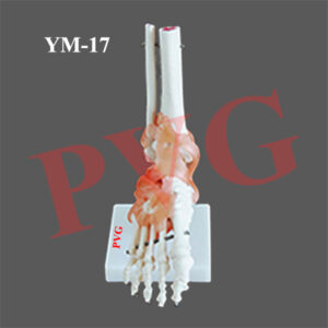
Anatomical Models
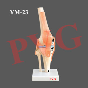
Anatomical Models
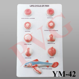
Anatomical Models
Anatomical Models
Anatomical Models
Anatomical Models

Anatomical Models

Anatomical Models
Reviews
There are no reviews yet.