
Be the first to review “YM-195 MODEL OF SEED GERMINATION”
Related products
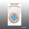
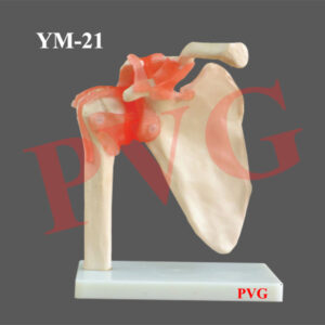
Anatomical Models
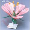
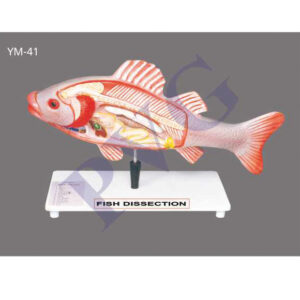
Anatomical Models
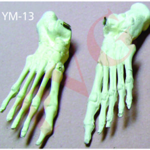
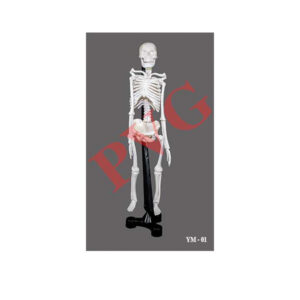
Anatomical Models

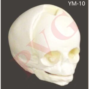
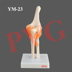
Anatomical Models
Anatomical Models

Anatomical Models
Anatomical Models
Anatomical Models
Reviews
There are no reviews yet.