
YM-198 MODEL OF DICOT AND MONOCOT ROOT

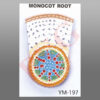
Transverse sectioned models of both mounted on one base board
for easy comparison of students. Size 12 x 24 x 3”.
Be the first to review “YM-198 MODEL OF DICOT AND MONOCOT ROOT”
Related products
Anatomical Models

Anatomical Models
Anatomical Models

Anatomical Models
Anatomical Models
YM-04 HUMAN SKELETON WITH NERVES & BLOOD VESSELS (TALL 85 cms)

Anatomical Models

Anatomical Models

Anatomical Models
Reviews
There are no reviews yet.